
Revolutionary 3D OCT - Optical Coherence Tomography.
We believe that seeing beneath the surface is essential to the health of your eyes, so we are absolutely delighted to offer state-of-the-art 3D Optical Coherence Tomography in both our Practices.
Revolutionary 3D OCT screening can instantly detect potentially serious conditions that can afffect your eyesight and your overall health, giving early diagnosis to a number of common ocular conditions.
How does OCT work?
- OCT is a new, completely painless, non-invasive, quick and highly advanced imaging system that checks for potentially serious eye conditions.
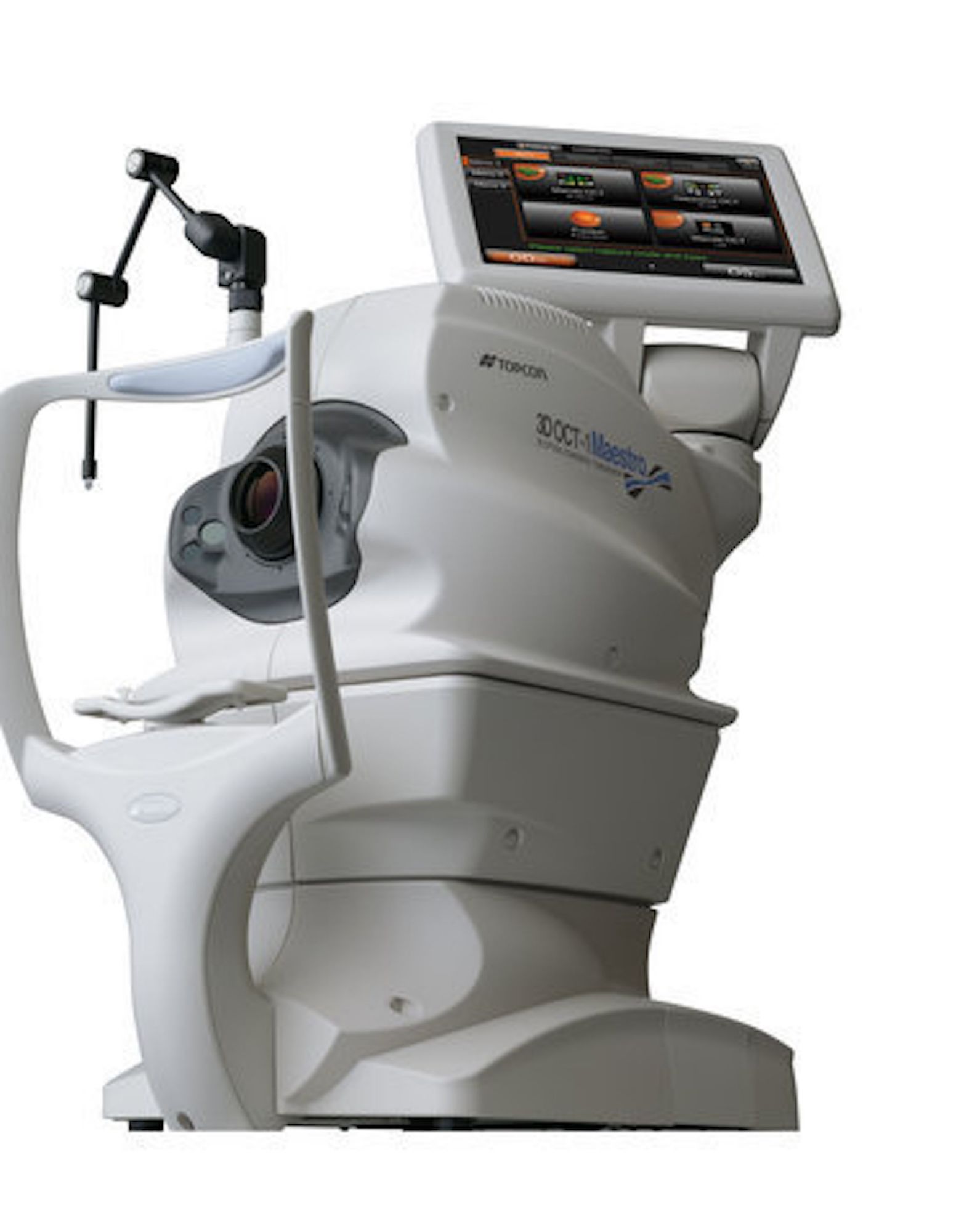
- Similar to ultrasound, OCT uses light rather than sound waves to image the different layers at the front and the back of the eye.
- The OCT machine captures both a digital photograph and a 3D cross sectional scan in one sitting.
What happens next?
- Make an appointment at one of our practices.
- Amanda will scan your eyes sing the 3D OCT camera.
- Your high resolution 3D images will be examined by amanda using specialist built-in analysis tools.
- Your results with be discussed with you.
- Your scans will be saved for comparative diagnosis.
What can the OCT scan check for?
Age-related Macular Degeneration
AMD is the leading cause of blindness in the UK. OCT can help to identify the earliest signs of AMD, determine whether it is the dry or the wet form and help monitor its progress over time.
Diabetes
Over 4 million people are now diagnosed with diabetes in the UK. An OCT examination helps enable early detection of diabetic retinopathy, allowing early referral and management which can greatly improve the success rate of treatment.
Glaucoma
Recent studies suggest that some form of glaucoma affects around 2 in 100 people over the age of 40, rising to almost 1 in 10 people over 75 years. OCT can facilitate early diagnosis so ongoing damage can be prevented and enable close monitoring to identify changes.
Vitreous Detachments
OCT provides invaluable information about the current relationship between the vitreous and the retinal surface of the eye. As we get older, the vitreous jelly that takes up the space in our eyeball can change, becomes less firm and can move away from the back of the eye towards the centre; in some cases parts do not detach and cause 'pulling' of the retinal surface. Quite often there is no pain and your eyesight will seem unchanged but the back of your eye may be being damaged.
Macular Holes
A macular hole is a small hole in the macular, the part of the retina responsible for our sharp, detailed central vision. Macular holes usually form during a complicated vitreous detachment when the vitreous pulls away from the back of the eye, causing a hole to form. Management of this condition needs to be carried out by and ophthalmologist in hospital.